
 Our Location

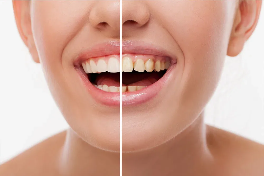
Restorative Dental Treatment: Medical Repair Processes for Cavities, Fractures, and Wear
The oral and dental structure plays a vital role not only as the first step of the nutritional process, but also in preserving phonetics (speech) and overall jaw anatomy. While many tissues in the human body have the capacity to renew themselves when damaged, the enamel tissue that forms the outermost layer of the tooth does not have such a cellular regeneration mechanism. Substance loss in the tooth due to decay, trauma, or structural wear can only be repaired through physician intervention using medical materials.
One of the most fundamental and commonly applied branches of dentistry, restorative dental treatment encompasses all clinical procedures aimed at restoring the anatomical form, chewing function, and biological integrity of damaged hard dental tissues (enamel and dentin). In this guide, the situations in which restorative treatments are necessary, clinical diagnostic methods, filling materials used, and scientific facts about the healing process are addressed in an objective manner.
What Is Restorative Dental Treatment and What Is Its Primary Purpose?
Restorative dental treatment is the process of compensating for superficial and moderately deep tissue losses that have not progressed to the tooth pulp (the inner chamber where the nerve and vascular bundle is located). If the infection reaches the tooth nerve, this falls not under restorative treatment but within the scope of endodontics (root canal treatment).
The primary medical goal of this treatment branch is to stop the destruction in the tooth and keep the tooth alive in the oral environment. By preserving the remaining healthy tooth tissue, the missing parts are completed with tissue-friendly (biocompatible) materials. In this way, the patient is able to grind food properly, unbalanced loads on the jaw joint are prevented, and the mutual bite relationship of the teeth (occlusion) is preserved.
In Which Cases Is Restorative Treatment Applied?
Whether a tooth requires restorative treatment is determined as a result of a detailed intraoral examination and radiological (X-ray) evaluation. The main clinical conditions in which this medical approach is applied are as follows:
1. Tooth Decay (Caries)
Bacteria present in the oral flora ferment the carbohydrate foods consumed and produce lactic acid. This acid dissolves the calcium and phosphate minerals in the tooth’s enamel layer (demineralization), forming decay cavities (holes). Cleaning the decay and filling the cavity is the most common situation encountered in restorative dentistry.
2. Physical Trauma and Fractures
As a result of falls, impacts, traffic accidents, or carelessly biting hard-shelled foods, chipping and fractures may occur in the crown portion of the tooth. Depending on the size of the fracture, this tissue loss is repaired with restorative materials in faithful accordance with the original form.
3. Tooth Wear (Attrition, Erosion, Abrasion)
The habit of clenching or grinding teeth during sleep (bruxism) leads to severe wear on the chewing surfaces of the teeth (attrition). Incorrect and forceful toothbrushing techniques create V-shaped notches (abrasion) in the cervical areas of the teeth, while frequent consumption of acidic drinks or reflux disease causes chemical dissolution of the enamel layer (erosion). All such structural wear is treated with restorative methods.
4. Closing Tooth Gaps (Diastema)
Gaps between the front teeth may lead to food impaction and, as a result, create a predisposition to gum infections (gingivitis). In cases where orthodontic treatment is not deemed appropriate, these anatomical spaces can be closed with restorative filling materials without damaging the tissue.
Medical Materials Used in Restorative Procedures
In modern medical science, the use of amalgam (commonly known as silver or black filling) has largely been abandoned due to its mercury content and aesthetic disadvantages. In current clinical practice, structures that can form a chemical bond with the tissue are preferred.
Composite Resins: These are light-cured, polymer-based materials containing inorganic fillers. They can be produced in shades compatible with the tooth’s natural color. Since they adhere to the tooth through a chemical bonding mechanism called "adhesion," there is no need for additional removal of healthy tooth tissue after the decay is cleaned.
Glass Ionomer Cements: They are generally used in pediatric patients, in the repair of primary teeth, or as a base material in very deep cavities to protect the tooth nerve. Thanks to their ability to release fluoride from within, they increase the tooth’s resistance to decay.
Indirect and Direct Restorations: Inlay and Onlay Systems
The technique applied changes depending on the extent of tissue loss in the tooth. Based on the clinical evaluation, physicians decide between direct or indirect treatment protocols.
Direct Restorations: Preferred in cases where the decay is not very extensive. The tooth is cleaned, and the prepared cavity is filled with composite material by the physician in a single session, layer by layer, in the clinic, and hardened with light of a specific wavelength. The treatment is completed the same day after polishing.
Indirect Restorations (Inlay and Onlay): Applied in cases where a large portion of the cusps on the chewing surface of the tooth has broken or decayed, but full coverage of the tooth (a crown) is not required. After the decay is removed in the clinic, a millimetric impression of the tooth is taken. In the laboratory, special fillings (inlay/onlay) are produced from porcelain or reinforced composite blocks, fitting the cavity in the tooth exactly like a puzzle piece. In the second session, this structure is permanently bonded to the tooth with medical cements.
Step-by-Step Clinical Workflow and Treatment Stages
The medical steps followed in a standard restorative treatment process are as follows:
Local Anesthesia: To prevent the patient from feeling pain while the decayed tissue is being removed, nerve conduction in the relevant area is temporarily blocked with anesthetic solutions.
Cavity Preparation: The physician cleans the infected (decayed) enamel and dentin tissue using special rotary instruments until healthy tissue is reached.
Isolation: The tooth to be treated must be protected from saliva and moisture. Composite materials are extremely sensitive to moisture. For this reason, the tooth is isolated with cotton rolls or rubber sheets called "rubber dam".
Acid and Bond Application: The tooth surface is roughened with medical acids, and "bond" (adhesive agent) is applied to ensure the filling adheres to the tooth.
Filling and Curing: The composite material is placed into the tooth in small increments. Each layer is hardened with halogen or LED devices.
Finishing and Polishing: After the filling is placed in the tooth, the occlusal height is checked. Surface irregularities are removed and the tooth is given its natural shine (polishing).
Oral Care and Follow-Up After Treatment
There is no rule that a tooth treated restoratively will never decay again. The material does not decay, but the boundary between the filling and the tooth may create an area of leakage for bacteria.
After treatment, food should not be consumed until the effect of anesthesia has completely worn off (approximately 2 hours) to prevent biting the lip or tongue.
Teeth should be brushed twice a day using the correct technique, and food trapped between the fillings must be cleaned with dental floss.
Cracking hard-shelled nuts such as walnuts and hazelnuts with the teeth can damage even the most durable fillings; such traumatic actions should be avoided.
Dental check-ups should be repeated every six months without fail.
Frequently Asked Questions
Will I feel a lot of pain during the filling procedure? Since the nerve network supplying the relevant tooth is numbed with local anesthesia before the procedure, no sharp pain is felt during the intervention. Only the vibration and pressure caused by the instruments used by the physician may be perceived.
Will tooth decay heal on its own if left untreated? Enamel tissue has no blood circulation or cellular regeneration activity. While a skin wound may form a scab and heal, a cavity that begins in the tooth will continue to progress unless medical intervention (mechanical cleaning and filling) is performed.
How long do composite fillings last? The clinical lifespan of fillings varies depending on the oral acid balance, the patient’s brushing habits, and dietary routine. With ideal oral care, restorations can function without problems for decades.
Legal Notice: This text has been prepared solely for the purpose of informing the public and increasing health literacy, in compliance with the regulations of the Regulation on Promotional and Informational Activities in Healthcare Services. The medical information presented here can in no way replace the clinical examination, diagnosis, or patient-specific treatment plan prepared by a dentist. For definitive diagnosis and medical guidance regarding your oral and dental health issues, please consult authorized healthcare institutions.